
Understanding orthodontic root resorption, advanced bite correction, and what the latest scientific research means for patients in Austin and Round Rock, Texas.
When considering braces, Invisalign, or other orthodontic treatments, patients and parents often have an important question: Can moving teeth affect the roots of those teeth?
It is an excellent question—and one that orthodontists take seriously.
Modern orthodontics is about much more than creating straight teeth. It involves understanding the relationship between teeth, roots, supporting bone, facial development, and the biological processes that make tooth movement possible.
A newly published scientific study co-authored by Dr. Anna Auladell of Bee Cave Orthodontics in Austin and Round Rock, Texas, provides important insight into one aspect of this relationship: orthodontically induced root resorption, or the shortening of tooth roots that can sometimes occur during orthodontic treatment.
Published in APOS Trends in Orthodontics in November 2026, the study examines how the roots of upper premolars and molars respond when orthodontists use specialized skeletal anchorage techniques to correct an anterior open bite.
The research is particularly interesting because it examines individual roots across an entire segment of the upper jaw rather than looking at just one or two teeth.
The findings offer valuable information about how different teeth respond to orthodontic forces, why root development matters, and how scientific research can help orthodontists make more informed treatment-planning decisions.
For patients considering orthodontic treatment in Austin, Bee Cave, Lakeway, Westlake, or Round Rock, this research provides another reason to look beyond cosmetic results when choosing an orthodontist.
1. Advancing the Science of Orthodontics
At Bee Cave Orthodontics, we believe exceptional orthodontic care combines clinical experience, advanced education, modern technology, and an ongoing commitment to understanding the science behind tooth movement.
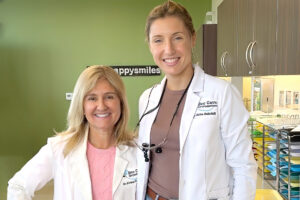
Dr. Anna Auladell brings an international educational background and a strong interest in orthodontic research to our practice.
Originally from Barcelona, Spain, Dr. Auladell completed her dental education at the Universitat de Barcelona and advanced orthodontic training at the Universitat Internacional de Catalunya. She subsequently completed additional orthodontic residency training at the Georgia School of Orthodontics in the United States.
She is a Diplomate of the American Board of Orthodontics and is a member of professional organizations including the American Association of Orthodontists.
Her research interests include orthodontic biomechanics, skeletal anchorage, and the treatment of complex bite problems.
This latest scientific contribution is especially relevant to patients because it examines a question that orthodontists must consider when planning certain types of tooth movement: How do the roots of teeth respond to the forces used to move them?
At Bee Cave Orthodontics, Dr. Auladell brings this research-oriented perspective to the care of children, teenagers, and adults seeking personalized orthodontic treatment.
2. Understanding Orthodontic Root Resorption: Can Braces Shorten Your Tooth Roots?
One of the lesser-known biological effects of orthodontic treatment is orthodontically induced root resorption (OIRR).
Root resorption occurs when the body breaks down and removes small amounts of the hard tissue that makes up a tooth root. In orthodontics, this process most commonly affects the tip, or apex, of the root.
To understand why this happens, it helps to understand how orthodontic treatment works.
Teeth are not permanently fixed in the jawbone. They are supported by a specialized structure called the periodontal ligament, which connects each tooth root to the surrounding bone.
When an orthodontist applies controlled pressure to a tooth, the supporting tissues respond through a biological process involving bone remodeling. Bone is gradually resorbed on one side of the tooth and formed on the other, allowing the tooth to move through the jaw.
This process is essential to orthodontic treatment.
However, the same mechanical forces that allow teeth to move can sometimes produce changes in the root structure.
These changes are known as orthodontically induced root resorption.
Is root resorption common with braces?
Small amounts of root resorption can occur during orthodontic treatment. The degree varies considerably among patients and even among individual teeth in the same patient.
Research has identified several factors that can influence the likelihood and extent of root resorption, including:
- The type and magnitude of orthodontic forces.
- The direction and duration of tooth movement.
- The type of tooth being moved.
- Individual root anatomy.
- Genetic and biological susceptibility.
- The maturity and developmental stage of the tooth root.
- The mechanics used to achieve a particular tooth movement.
Importantly, root resorption is not exclusive to traditional braces. It is a biological response that can occur with different types of orthodontic tooth movement.
It is also important to understand that not every patient experiences clinically significant root resorption.
A systematic review by Bellini-Pereira and colleagues, published in the European Journal of Orthodontics in 2021, evaluated root resorption following orthodontic intrusion and highlighted the importance of understanding the relationship between orthodontic mechanics and biological response.
The study by Dr. Auladell and her colleagues adds to this research by examining the response of multiple individual roots following posterior intrusion using miniplates.
3. What Was the New Study About?
Study title: Root Length Changes in the Maxillary Posterior Segment Following Miniplate-Anchored Intrusion: A Retrospective Cone-Beam Computed Tomography Study.
Journal: APOS Trends in Orthodontics.
Publication: Article in press, November 11, 2026.
DOI: 10.25259/APOS_324_2026.
Study design: Retrospective observational cohort study.
Sample size: 19 patients.
Primary objective: To evaluate changes in the roots of upper premolars and molars following miniplate-anchored orthodontic intrusion.
The study focused on a particular orthodontic movement called posterior intrusion.
Posterior intrusion involves moving the upper back teeth vertically into the jawbone. This technique can be used to help correct an anterior open bite, a condition in which the upper and lower front teeth do not make contact when the back teeth are together.
Correcting this type of bite problem can require sophisticated orthodontic biomechanics.
Rather than relying exclusively on conventional braces or aligners, orthodontists may use temporary skeletal anchorage devices to provide a stable point from which to apply controlled forces.
The researchers wanted to understand how the different roots of the upper premolars and molars responded to this treatment.
Who conducted the research?
The study was conducted by six researchers:
- Anna Auladell – Department of Orthodontics, Georgia School of Orthodontics, Atlanta, Georgia, USA.
- Prasad Nalabothu – Department of Pediatric Oral Health and Orthodontics, University Center for Dental Medicine UZB, Basel, Switzerland.
- Hayden Wynne – Department of Orthodontics, Georgia School of Orthodontics, Atlanta, Georgia, USA.
- Niki Zoka – Department of Orthodontics, Georgia School of Orthodontics, Atlanta, Georgia, USA.
- Courtney McCracken – Medalytics Group, Northridge Plaza, Atlanta, Georgia, USA.
- Nandakumar Janakiraman – Department of Orthodontics, Georgia School of Orthodontics, Atlanta, Georgia, USA.
Dr. Anna Auladell and Prasad Nalabothu were designated joint first authors and contributed equally to the work.
Nandakumar Janakiraman was the corresponding author.
Recognizing all six researchers is important because this was a collaborative scientific investigation involving orthodontic expertise, imaging analysis, and research methodology.
4. What Are Miniplates, and Why Do Orthodontists Use Them?
One of the most interesting aspects of this study is the orthodontic technique being investigated.
Miniplates are small titanium anchorage devices that are temporarily attached to the jawbone. They provide a stable point from which orthodontists can apply forces to move teeth.
They are part of a broader category of treatment techniques known as skeletal anchorage.
Traditional orthodontic mechanics often rely on other teeth to provide anchorage. For example, moving one group of teeth may require using another group as a stabilizing point.
Skeletal anchorage provides an alternative by using the supporting bone rather than relying exclusively on other teeth.
How does miniplate-anchored intrusion work?
In the study, titanium miniplates were placed on both sides of the upper jaw in an area called the infrazygomatic crest.
The plates were secured with small screws and used as anchorage points for the orthodontic forces needed to intrude the upper posterior teeth.
This technique offers several potential advantages in appropriately selected cases:
- Provides a stable anchorage point.
- Allows controlled vertical movement of posterior teeth.
- Reduces dependence on other teeth for anchorage.
- Can help orthodontists manage more complex open-bite mechanics.
- Requires less reliance on patient compliance for the anchorage component.
Miniplates are not necessary for most orthodontic patients. They are specialized devices used when the treatment objectives and biomechanics warrant them.
Their use is particularly relevant in certain patients with anterior open bites, vertical skeletal discrepancies, or complex posterior tooth movements.
The research by Dr. Auladell and her colleagues specifically examined the biological response to this technique.
5. How Did the Researchers Measure Changes in Tooth Roots?
A major strength of the study was its use of three-dimensional cone-beam computed tomography, commonly known as CBCT.
CBCT imaging provides a three-dimensional view of the teeth and surrounding structures.
Unlike conventional two-dimensional dental images, CBCT allows researchers to examine individual roots from multiple directions and identify anatomical landmarks with greater spatial detail.
For this study, researchers examined CBCT scans taken at two different times:
- T0: Before treatment.
- T1: Following completion of the posterior intrusion phase.
The researchers evaluated 19 patients who underwent miniplate-supported posterior intrusion to correct an anterior open bite.
The average interval between scans was approximately seven months, with a range of 4.5 to 8.5 months.
They measured the roots of:
- Upper first premolars.
- Upper second premolars.
- Upper first molars.
- Upper second molars.
Each root was evaluated individually.
The measurements extended from a standardized reference point in the central fossa of the tooth to the tip of the corresponding root.
The researchers also used two independent examiners to evaluate measurement reliability. Their intraclass correlation coefficients exceeded 0.76, indicating strong agreement.
This methodology allowed the research team to identify differences in root response that might otherwise be difficult to appreciate using conventional imaging.
An important qualification: The study evaluated linear root-length changes, not the total volume of root tissue lost.
6. What Did the Researchers Discover? The Most Important Findings
The findings demonstrated that not all teeth respond to orthodontic intrusion in the same way.
Some roots experienced measurable shortening, while others showed little change or continued development.
The greatest changes occurred in the premolars and first molars. The second molars, which included teeth with incompletely developed roots, showed no statistically significant root shortening.
Here is a closer look at the results.
Finding No. 1: Upper premolars experienced the greatest shortening in certain roots
The researchers found measurable root shortening in several upper premolars.
The greatest average change occurred in the palatal root of the upper right first premolar, which showed a mean reduction of 0.99 millimeters.
Other significant findings included:
- Upper left second premolar, palatal root: 0.76 mm reduction.
- Upper left first premolar, palatal root: 0.47 mm reduction.
- Upper left first premolar, buccal root: 0.66 mm reduction.
- Upper right second premolar, palatal root: 0.51 mm reduction.
The researchers found that palatal roots were particularly susceptible to changes during this type of intrusion.
Finding No. 2: First molars also experienced measurable root shortening
The first molars demonstrated a similar pattern.
The researchers identified the greatest average shortening in the palatal and distobuccal roots.
For the upper right first molar:
- Palatal root: 0.98 mm reduction.
- Distobuccal root: 0.96 mm reduction.
- Mesiobuccal root: 0.53 mm reduction.
For the upper left first molar:
- Palatal root: 0.47 mm reduction.
- Distobuccal root: 0.75 mm reduction.
- Mesiobuccal root: No statistically significant change.
These findings demonstrate why examining individual roots can be valuable.
A molar has multiple roots, and those roots do not necessarily experience identical mechanical forces during orthodontic movement.
Finding No. 3: Second molars showed no statistically significant root resorption
Perhaps one of the most interesting findings involved the upper second molars.
Unlike the premolars and first molars, the second molars did not demonstrate statistically significant root shortening.
In fact, the researchers observed small increases in measured root length.
For example:
- Upper right second molar: increases ranging from 0.02 to 0.09 mm.
- Upper left second molar: increases ranging from 0.31 to 0.49 mm.
None of these changes reached statistical significance.
The researchers suggested that continued root development may explain these findings.
Some second molars had incompletely formed root apices at the beginning of treatment. These developing roots may have different biological responses to orthodontic forces than fully mature roots.
This observation reinforces an important consideration in orthodontic treatment planning: the developmental stage of a tooth can matter.
Summary of the study’s principal findings
| Tooth group | Principal finding |
|---|---|
| Upper premolars | Significant root shortening in most evaluated roots |
| Upper first molars | Significant shortening in several roots, especially palatal and distobuccal |
| Upper second molars | No statistically significant root shortening |
| Greatest measured change | 0.99 mm in the upper right first premolar’s palatal root |
| Treatment interval | Approximately 7 months on average |
The researchers concluded that miniplate-anchored intrusion was associated with limited, less-than-1-mm root resorption in the premolars and first molars examined.
They characterized these changes as clinically acceptable within the context of their study.
That conclusion should be interpreted within the limits of this particular research rather than as a guarantee that every patient will experience the same result.
7. Why Does the Age and Development of a Tooth Matter?
One of the most meaningful clinical implications of this study is the relationship between root development and orthodontic root resorption.
Teeth do not all finish developing at the same time.
In children and teenagers, some permanent teeth may still be developing even after they have erupted into the mouth.
The researchers observed that second molars with developing roots appeared less susceptible to measurable apical root shortening during the period examined.
Why might this happen?
Developing roots have different biological characteristics than fully mature roots.
The study discusses several possible explanations:
- Greater cellular activity.
- Increased reparative capacity.
- A wider opening at the root apex during development.
- Differences in how mechanical stress is distributed.
These biological differences may help explain why developing roots can respond differently to orthodontic forces.
For parents considering orthodontic treatment for their children, this is another reason why an individualized evaluation matters.
The appropriate timing of orthodontic treatment depends on the child’s dental development, jaw growth, bite relationship, and specific treatment needs.
It is not simply a matter of starting braces as early as possible.
A qualified orthodontist evaluates these factors to determine when intervention may be beneficial and which treatment approach is appropriate.
8. Why Three-Dimensional Imaging Matters in Modern Orthodontics
Another important contribution of this research is its use of CBCT imaging.
Traditional dental radiographs provide valuable diagnostic information, but two-dimensional images have inherent limitations.
Roots can overlap, anatomical structures can obscure important details, and image magnification can affect measurements.
Three-dimensional CBCT imaging allows orthodontists and researchers to examine the spatial relationships of teeth and roots from different perspectives.
In this particular study, the researchers used CBCT to compare root lengths before and after treatment.
This helped them identify patterns that might be difficult to evaluate using conventional imaging alone.
However, an important distinction must be made.
CBCT is not necessary for every orthodontic patient.
Three-dimensional imaging involves additional radiation exposure compared with conventional dental imaging. It should be used when the diagnostic benefit justifies the exposure and when the information is expected to contribute meaningfully to treatment planning.
The goal of modern orthodontics is not to take more images. It is to use the appropriate diagnostic tools for each patient’s needs.
At Bee Cave Orthodontics, advanced digital scanning and individualized treatment planning are part of our approach to evaluating patients and developing treatment strategies.
9. What Does This Research Mean for Someone Considering Braces or Invisalign?
Patients frequently ask whether braces or Invisalign can damage their teeth.
The honest answer is that orthodontic treatment involves biological changes, and root resorption is one potential risk that orthodontists consider.
However, the research by Dr. Auladell and her colleagues should not be interpreted as evidence that all braces or Invisalign treatments cause the same degree of root shortening.
There are several important distinctions.
First, this study evaluated a specific treatment technique.
It examined miniplate-anchored intrusion of upper posterior teeth. It did not compare Invisalign with traditional braces, nor did it evaluate every type of orthodontic movement.
Second, root resorption varies from patient to patient.
Individual anatomy, tooth development, biological susceptibility, and treatment mechanics all play a role.
Third, the study reinforces the importance of treatment planning.
Understanding the direction, magnitude, and duration of orthodontic forces can help clinicians evaluate potential risks when planning complex movements.
Fourth, the findings do not suggest that patients should avoid orthodontic treatment.
Rather, they provide additional information that orthodontists can consider when evaluating the potential benefits and risks of treatment.
The ultimate objective is to create a healthy, functional bite and an attractive smile while protecting the long-term health of the teeth and supporting structures.
10. What Is an Anterior Open Bite, and How Do Orthodontists Correct It?
Because the research focused on patients with anterior open bites, it is worth explaining this condition.
An anterior open bite occurs when the upper and lower front teeth do not touch when the back teeth are brought together.
Depending on the cause and severity, an open bite may affect:
- Biting into foods.
- Speech and pronunciation.
- The appearance of the smile.
- The relationship between the upper and lower jaws.
- Overall dental function.
Open bites can develop for several reasons, including skeletal growth patterns, dental positioning, tongue habits, and other contributing factors.
Treatment depends on identifying the underlying cause.
Some patients may benefit from conventional orthodontic treatment. Others may require more advanced mechanics, skeletal anchorage, or collaboration with an oral surgeon.
Posterior intrusion is one approach used in selected cases.
By moving the upper back teeth vertically, orthodontists may help allow the lower jaw to rotate counterclockwise, improving the relationship between the upper and lower front teeth.
Miniplates can provide the anchorage needed to accomplish this type of movement in appropriately selected patients.
The research by Dr. Auladell and her colleagues contributes to understanding the biological response associated with this particular treatment approach.
11. How Bee Cave Orthodontics Combines Research, Experience, and Personalized Treatment
Scientific research is valuable because it helps orthodontists better understand how treatment works.
But research is only one part of providing excellent patient care.
At Bee Cave Orthodontics, our philosophy combines specialized orthodontic knowledge, individualized treatment planning, advanced technology, and an understanding that every patient has different needs.
Our team includes two orthodontists with distinctive professional backgrounds.
Dr. Anna Auladell, DDS
Dr. Auladell brings international education, specialized orthodontic training, and published research experience to Bee Cave Orthodontics.
Her scientific work examines orthodontic biomechanics and the biological changes that can occur when teeth are moved.
Her co-authorship of the 2026 miniplate intrusion study reflects her commitment to advancing the scientific understanding of orthodontic treatment.
She is also a Diplomate of the American Board of Orthodontics.
Dr. Adriana Da Silveira, DDS, MS, PhD
Dr. Adriana Da Silveira brings more than 25 years of orthodontic experience, advanced academic education, and extensive experience treating children, teenagers, and adults.
Her educational background includes a PhD in Oral Biology, a Master of Science, and specialized orthodontic training.
She also has extensive experience treating complex orthodontic and craniofacial cases.
Together, our doctors bring complementary educational backgrounds and clinical perspectives to the practice.
What patients can expect at Bee Cave Orthodontics
Our practice provides orthodontic care for children, teenagers, and adults, including:
- Traditional braces.
- Invisalign clear aligners.
- Invisalign First for younger patients.
- Early orthodontic evaluations.
- Treatment for complex bite problems.
- Personalized orthodontic treatment planning.
- Digital 3D scanning.
We believe patients deserve to understand their treatment options, the reasoning behind their treatment recommendations, and the factors that can influence their long-term results.
Our goal is to make orthodontic care accessible, understandable, and tailored to each individual.
12. Choosing an Orthodontist in Austin or Round Rock: Why Experience and Research Matter
Choosing an orthodontist is an important decision for individuals and families.
Whether you are considering braces for your child, Invisalign for yourself, or treatment for a more complicated bite problem, it is important to understand the qualifications and experience of the doctor overseeing your care.
Here are several questions worth considering when selecting an orthodontist:
Is the doctor a specialist in orthodontics?
Orthodontists complete additional specialized education beyond dental school to diagnose and treat problems involving tooth alignment, jaw relationships, and bite development.
Does the doctor have experience treating complex cases?
Not every orthodontic problem is simply a matter of straightening teeth. Some require advanced biomechanics and an understanding of skeletal growth and facial structure.
Does the practice use modern diagnostic technology?
Digital scanning, appropriate imaging, and careful treatment planning can help orthodontists evaluate individual needs and monitor progress.
Does the orthodontist explain potential risks and benefits?
Patients should understand the treatment process, including possible biological changes such as root resorption.
Does the treatment plan reflect the individual patient?
No two patients have exactly the same dental anatomy, growth patterns, or treatment objectives.
At Bee Cave Orthodontics, we believe these considerations are essential to delivering responsible, high-quality orthodontic care.
We welcome families from Austin, Bee Cave, Lakeway, Westlake, Spicewood, Round Rock, and surrounding Central Texas communities.
Frequently Asked Questions About Orthodontic Root Resorption
These are some of the questions patients commonly ask when researching orthodontic treatment, root health, and bite correction.
1. Can braces cause tooth roots to shorten?
Yes. Orthodontic treatment can sometimes cause a process called orthodontically induced root resorption, in which small amounts of root tissue are lost, most commonly near the root tip. The amount varies among patients and teeth. Many changes are small, but more significant root resorption can affect long-term tooth prognosis. Orthodontists consider this possibility when planning treatment.
2. Does Invisalign cause root resorption?
Root resorption is a biological response to orthodontic tooth movement, not a complication exclusive to traditional braces. It can potentially occur with different orthodontic appliances, including clear aligners. The 2026 study by Auladell and colleagues specifically investigated miniplate-anchored intrusion, not Invisalign treatment.
3. What is orthodontic root resorption?
Orthodontic root resorption is the loss of some of the hard tissue that makes up a tooth root during orthodontic treatment. It most commonly occurs at the root apex. Its severity depends on multiple biological and mechanical factors.
4. Is root resorption from braces permanent?
Root tissue that has been lost through resorption does not regenerate in the same way as ordinary bone remodeling. However, orthodontic forces can be adjusted or paused when clinically appropriate to manage risk. The significance of root shortening depends on the amount of tissue lost, the tooth involved, and the patient’s overall dental health.
5. How much root shortening is considered acceptable?
There is no single measurement that is universally acceptable for every patient. The clinical significance depends on the original root length, the extent and location of resorption, the health of the surrounding structures, and other individual factors.
In the 2026 study by Dr. Auladell and colleagues, the observed root shortening was less than 1 mm in the evaluated premolars and first molars. The authors characterized these changes as clinically acceptable in the context of their study.
6. Can orthodontists detect root resorption?
Orthodontists can evaluate potential root changes through appropriate dental imaging and clinical assessment. Depending on the situation, conventional radiographs or three-dimensional CBCT imaging may be considered. CBCT is not routinely necessary for every patient.
7. Does age affect the risk of root resorption?
Tooth development can influence the biological response to orthodontic forces. The Auladell study found that second molars with developing roots showed no statistically significant root shortening during the study period. However, this does not establish that younger patients are universally protected from root resorption.
8. What are miniplates in orthodontics?
Miniplates are small titanium devices temporarily attached to the jawbone. They provide stable anchorage for orthodontic forces and can help move teeth in directions that may be difficult to accomplish using conventional anchorage alone.
9. What is posterior intrusion in orthodontics?
Posterior intrusion is the process of moving the upper or lower back teeth vertically into the supporting jawbone. It is sometimes used to help correct an anterior open bite by changing the vertical relationship of the back teeth and allowing the lower jaw to rotate.
10. Can an anterior open bite be corrected without jaw surgery?
Some anterior open bites can be corrected with orthodontic treatment alone. Others, particularly those involving significant skeletal discrepancies, may require a combination of orthodontics and orthognathic surgery. Treatment depends on the patient’s age, anatomy, growth, and the underlying cause of the open bite.
11. Should every orthodontic patient have a CBCT scan?
No. CBCT imaging should be prescribed when the additional three-dimensional information is expected to provide meaningful diagnostic or treatment-planning benefits. It is not necessary for every orthodontic patient.
12. Who is Dr. Anna Auladell?
Dr. Anna Auladell is an orthodontist at Bee Cave Orthodontics, serving patients in Austin and Round Rock, Texas. Originally from Barcelona, Spain, she completed dental and orthodontic education in Spain and specialized orthodontic residency training in Georgia. She is a Diplomate of the American Board of Orthodontics and a co-author of peer-reviewed research involving orthodontic biomechanics and root changes following posterior intrusion.
13. Where can I find an orthodontist in Austin or Round Rock, Texas?
Bee Cave Orthodontics offers specialized orthodontic care at two Central Texas locations:
- Austin/Bee Cave: 11412 Bee Cave Road, Suite 300, Austin, TX 78738.
- Round Rock: 1499 E. Old Settlers Boulevard, Round Rock, TX 78664.
The practice provides orthodontic treatment for children, teenagers, and adults, including braces and Invisalign.
14. Does Bee Cave Orthodontics offer consultations?
Yes. Bee Cave Orthodontics welcomes new patients and offers consultations to help individuals and families understand their orthodontic needs and explore treatment options.
The Bottom Line: Better Orthodontics Starts With Understanding the Science
Orthodontics is both an art and a science.
Creating a beautiful smile requires much more than moving teeth into alignment. It requires understanding how teeth, roots, supporting bone, and the jaws respond to carefully controlled forces.
The research co-authored by Dr. Anna Auladell provides valuable insight into the biological response of different teeth following miniplate-anchored intrusion.
By examining individual roots across the upper posterior segment, the researchers demonstrated that root resorption is not uniform. Premolars and first molars experienced measurable shortening, while second molars with developing roots showed no statistically significant resorption during the observation period.
The study also reinforces the importance of considering root development, orthodontic mechanics, and individual biological differences when planning treatment.
For patients and families in Austin and Round Rock, the message is straightforward: choosing an orthodontist who understands both the science and the clinical practice of tooth movement matters.
At Bee Cave Orthodontics, Dr. Anna Auladell, Dr. Adriana Da Silveira, and our team are committed to providing individualized orthodontic care grounded in education, experience, and an understanding of the science behind every smile.
Ready to learn more about your orthodontic options?
Whether you are considering braces, Invisalign, early orthodontic treatment for your child, or help correcting a more complex bite problem, we invite you to meet our team.
Bee Cave Orthodontics – Austin & Round Rock, Texas
Visit beecaveortho.com to request a consultation.
Scientific References and Further Reading
The following publications include the original research and supporting scientific literature cited by Auladell and colleagues. They provide additional context on orthodontic root resorption, intrusion mechanics, skeletal anchorage, and diagnostic imaging.
Primary research article
- Auladell A, Nalabothu P, Wynne H, Zoka N, McCracken C, Janakiraman N. Root length changes in the maxillary posterior segment following miniplate-anchored intrusion: A retrospective cone-beam computed tomography study. APOS Trends in Orthodontics. 2026. DOI: 10.25259/APOS_324_2026.
Supporting peer-reviewed research
- Bellini-Pereira SA, Almeida J, Aliaga-Del Castillo A, Dos Santos CC, Henriques JF, Janson G. Evaluation of root resorption following orthodontic intrusion: A systematic review and meta-analysis. European Journal of Orthodontics. 2021;43:432–441.
- Mauès CP, Do Nascimento RR, Vilella OD. Severe root resorption resulting from orthodontic treatment: Prevalence and risk factors. Dental Press Journal of Orthodontics. 2015;20:52–58.
- Al-Qawasmi RA, Hartsfield JK, Everett ET, Flury L, Liu L, Foroud TM, et al. Genetic predisposition to external apical root resorption. American Journal of Orthodontics and Dentofacial Orthopedics. 2003;123:242–252.
- Jung YH, Cho BH. External root resorption after orthodontic treatment: A study of contributing factors. Imaging Science in Dentistry. 2011;41:17–21.
- Guo Y, He S, Gu T, Liu Y, Chen S. Genetic and clinical risk factors of root resorption associated with orthodontic treatment. American Journal of Orthodontics and Dentofacial Orthopedics. 2016;150:283–289.
- Pandis N, Nasika M, Polychronopoulou A, Eliades T. External apical root resorption in patients treated with conventional and self-ligating brackets. American Journal of Orthodontics and Dentofacial Orthopedics. 2008;134:646–51.
- Costopoulos G, Nanda R. An evaluation of root resorption incident to orthodontic intrusion. American Journal of Orthodontics and Dentofacial Orthopedics. 1996;109:543–548.
- Han G, Huang S, Von Den Hoff JW, Zeng X, Kuijpers-Jagtman AM. Root resorption after orthodontic intrusion and extrusion: An intraindividual study. The Angle Orthodontist. 2005;75:912–918.
- Akl HE, El-Beialy AR, El-Ghafour MA, Abouelezz AM, El Sharaby FA. Root resorption associated with maxillary buccal segment intrusion using variable force magnitudes. The Angle Orthodontist. 2021;91:733–742.
- Al-Falahi B, Hafez AM, Fouda M. Three-dimensional assessment of external apical root resorption after maxillary posterior teeth intrusion with miniscrews in anterior open bite patients. Dental Press Journal of Orthodontics. 2018;23:56–63.
- Li W, Chen F, Zhang F, Ding W, Ye Q, Shi J, et al. Volumetric measurement of root resorption following molar mini-screw implant intrusion using cone beam computed tomography. PLoS One. 2013;8:e60962.
- Tarraf NE, Darendeliler MA. Miniscrews vs. miniplates. In: Temporary Anchorage Devices in Clinical Orthodontics. Wiley; 2020. p. 477–488.
- Omidkhoda M, Bardideh E, Jahanbin A, Zarei M. Effects of posterior intrusion using skeletal anchorage on treating anterior open bite: A systematic review and meta-analysis. Journal of Dental Research, Dental Clinics, Dental Prospects. 2023;17:196–210.
- Alves A, Cacho A, San Roman F, Gerós H, Afonso A. Mini implants osseointegration, molar intrusion and root resorption in Sinclair minipigs. International Orthodontics. 2019;17:733–743.
- St Martin JG, Javed F, Rossouw PE, Michelogiannakis D. Influence of mini-screw implant-assisted intrusion on orthodontically induced inflammatory root resorption: A systematic review. European Archives of Paediatric Dentistry. 2021;22:341–349.
- Ari-Demirkaya A, Masry MA, Erverdi N. Apical root resorption of maxillary first molars after intrusion with zygomatic skeletal anchorage. The Angle Orthodontist. 2005;75:761–767.
- Abu Arqub S, Al-Moghrabi D, Iverson MG, Farha P, Alsalman HA, Uribe F. Assessment of the efficacy of various maxillary molar intrusion therapies: A systematic review. Progress in Orthodontics. 2023;24:37.
- Xun C, Zhao H, Zeng X, Wang X. Intrusion of overerupted maxillary molars with miniscrew implant anchorage: A radiographic evaluation. Journal of Huazhong University of Science and Technology. 2013;33:780–785.
- Alamadi E, Alhazmi H, Hansen K, Lundgren T, Naoumova J. A comparative study of cone beam computed tomography and conventional radiography in diagnosing the extent of root resorptions. Progress in Orthodontics. 2017;18:37.
- Baena-De La Iglesia T, Yañez-Vico RM, Iglesias-Linares A. Diagnostic performance of cone-beam computed tomography to diagnose in vivo/in vitro root resorption: A systematic review and meta-analysis. Journal of Evidence-Based Dental Practice. 2023;23:101803.
About this article: This educational article is based primarily on the peer-reviewed research by Auladell, Nalabothu, Wynne, Zoka, McCracken, and Janakiraman, with additional background from the references cited in that study. It is intended for general educational purposes and is not a substitute for an individual orthodontic evaluation. Treatment recommendations and imaging decisions should be based on each patient’s clinical circumstances.